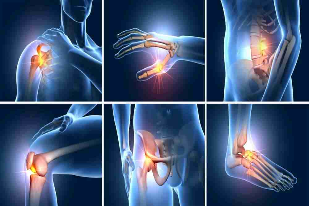
Osteoarthritis In All Body Regions

Osteoarthritis can affect multiple joints throughout the body. According to gender and other causal factors, the ratio of incidence may vary. In addition, there are minor variations in radiological findings and clinical manifestations. We can examine them individually:
Knee
The knee is one of the joints most commonly afflicted by osteoarthritis. According to a report, 37% of patients older than 60 years had knee osteoarthritis. It is also somewhat more prevalent in females. Due to its anatomical location and physiological function, the knee joint is most susceptible to osteoarthritis. It carries the weight of the entire trunk and upper limbs, and its stability is essential for the correct functioning of the lower limbs.
Abnormal knee alignments, such as in ‘varus’ (bow legs) and ‘valgus’ (knock knees), can also increase the likelihood of developing osteoarthritis. Varus alignment increases the likelihood of knee osteoarthritis on the medial side. Lateral OA is predisposed by valgus alignment. Again, this is due to enhanced force of action passing on successive sides.
Radiographs in the anterior-posterior and lateral positions can help detect aberrant osteoarthritis symptoms. OA can affect either the joint between the thigh bone and the leg bone or the patella, the knee cap. Typically, the joint between the patella and femur is afflicted first.
Hip
The hip joint is almost the second most frequently afflicted by osteoarthritis. A study reveals that 27 percent of patients older than 45 have hip joint OA. Between the apex of the large femur bone and its socket in the pelvic bone, the hip joint is formed. OA of the hip joint may cause discomfort in the area around the groin, which may radiate to the back and knee due to a loss of support at the trunk-lower body junction.
The osteoarthritis alterations are visible on a frontal plain x-ray. Most commonly, the upper and outer side of the joint bearing the most pressure and weight are affected. There are osteophytes or bony outgrowths at this location. Additionally, bone can be deformed at this site.
Fingers and hands
The hand is also a common site of arthritic inflammation. Approximately 27% of OA patients have the condition in their wrists. The most commonly affected joints in the hand are the joint where the thumb connects the wrist and the joints in the fingers. In the digits, distal joints closer to the nail bed may become afflicted, resulting in the formation of Heberden’s nodes. The middle joint of the finger is the source of Bouchard’s nodes. These nodes are enlargements caused by bony outgrowths or spurs as a result of cartilage degeneration in osteoarthritis.
Unlike osteoarthritis of other joints, osteoarthritis of the hands restricts the patient’s delicate motor skills. The patient will struggle with tasks such as buttoning garments, writing for extended periods, typing, and drawing.
As stated previously, the nodes cause the digits to become trembling and rigid. Similarly, if there are bony protrusions on the bones that unite to form the wrist, this can lead to carpal tunnel syndrome. The bony projections in the vicinity of these joints compress the nerves. This can cause hand agony comparable to being pricked by needles. The symptoms of decreased wrist motility include diminished grip strength, making it impossible to perform a variety of duties, such as holding a paintbrush or utensils, etc.
Neck, spine, and back
The joints in the spinal column are most frequently affected by osteoarthritis in old age, in accordance with the general trend in the incidence of the condition. Vertebra are the constituent bones that make up the spinal column. Osteoarthritis most commonly affects the facet joints between these vertebrae.
As this bony column protects the spinal cord, which is an extension of the brain, the effects of osteoarthritis of the spine may be more worrisome. Through passageways between these bones, nerves exit the vertebral canal. If these bones are compromised by osteoarthritis, the irregularities and spurs of the bones can compress the nerves that supply distant body regions. Consequently, the symptoms can range from localized pain in the neck, back, or lower spine when twisting, bending, or straightening, to weakness, numbness, and sensory loss in the extremities and other body parts. These additional symptoms will increase the disease’s impact on quality of life.
Radiographic findings may reveal deterioration of the vertebral bone blocks, a decrease in the articular cartilage between these bones resulting in bone fusion, and the presence of bone spurs. MRIs can also be utilized to detect spinal cord compression.
The shoulder
The shoulder joint is afflicted less frequently than the aforementioned joints. It is more likely to occur in sports involving overhead movements, direct impacts, or shoulder joint accidents. The shoulder joint consists of the humeral head in its cavity in the shoulder bone. The bone deformations associated with progressive osteoarthritis may result in the loss of normal ball and socket anatomy.
The axillary view of the radiographs accurately demonstrates any changes in the joint space, any swelling of the joint capsule, and may also rule out shoulder dislocation as a probable cause of pain.
The calf and foot
The big toe is by far the most common site of osteoarthritis in the foot. It can be caused by forceful kicking, falling objects, or twisting and bending joints at unnatural angles.
The complex picture of osteoarthritis in the great toe can lead to Hallux valgus, which is the inward turning of the great toe caused by bony outgrowth on the lateral side of the toe. This protrusion is also known as a bunion. Complete deterioration of the cartilage in this joint can lead to a condition known as Hallux rigidus.
If we trace a line of balance from the trunk to the ground, the ankle joint is the lowest point bearing pressure. The tendons at this joint may deteriorate, placing additional stress on the joint and rendering the patient immobilized and unable to walk. Shoes that position the feet at unnatural angles, such as sandals and high heels, can increase the risk of osteoarthritis in the feet.
