How Is Osteoarthritis Diagnosis Made?

A timely and accurate diagnosis of a disease is the first step in preventing the disease from progressing to a more serious condition. An orthopedic physician diagnoses osteoarthritis based on a number of criteria.
The first thing a doctor does to investigate a patient’s complaints is to obtain a complete and detailed medical history. The history includes inquiries regarding the complaints. The patient’s detailed responses provide the physician with an understanding of all the potential diseases the patient may be suffering from. Under the following headings, we can see how the physician conducts his or her investigations in order to diagnose osteoarthritis.
The clinical
Clinical investigation involves investigating the symptoms of osteoarthritis, such as joint discomfort and stiffness. The clinician may proceed by asking the following types of questions:
The onset, duration, and progression of pain, as well as whether or not the pain is made worse by inactivity or movement.
Any complaint of joint stiffness, particularly in the morning.
If the patient has observed any restricted mobility, movement limitations, etc., or any increase in work-related difficulties that can be attributed to the symptoms.
Any additional symptoms such as body pains, fever, heart palpitations, etc. These assist in ruling out alternative causes of joint discomfort, such as rheumatoid arthritis, gout, knee injury, etc.
Any joint injury in the past.
Any occurrence of comparable complaints among blood relatives, occupational history, etc.
Any specific dietary regimen the patient is following.
The physician will then inspect the joint. Any indications of joint capsule tenderness or enlargement will be noted. On the basis of these findings, additional investigations can be requested.
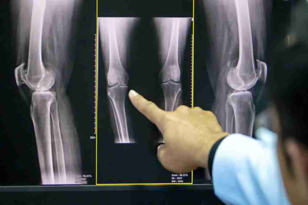
X-rays are the most common initial diagnostic tool for bone and joint diseases. A conventional film radiograph of the joint can reveal osteoarthritis symptoms. However, the physician may prescribe multiple x-ray views to better assess the situation. The following indications are visible on the radiograph:
If the joint space is diminished, it is a negative sign that can contribute to sclerosis of the bone ends.
The presence of bone spurs or protrusions at the joint capsule.
Any joint capsule enlargement, which may indicate early inflammation.
From time to time, multiple radiographs may be prescribed for accurately tracking the progression of the disease. Not always do radiographic findings correspond to clinical symptoms. The patient may report a worsening of symptoms, but the radiographs may not reveal a corresponding and significant deterioration of the structures.
The following is a classification of osteoarthritis:
The Kellgren and Lawrence system classifies osteoarthritis based on radiographic imaging as follows:
Grade 0 denotes the absence of any radiological indications
The image in Grade 1 is ambiguous.
Grade 2 demonstrates slight, but distinct indications of bony spur and/or joint space constriction.
Grade 3 displays moderate osteophyte formation, joint space narrowing, and skeletal deformity.
Grade 4 is distinguished by the presence of severe bony spurs, joint space sclerosis, and bone deformity.
Blood and joint fluid samples may be taken to rule out other conditions with similar symptoms.
