Evaluation Of Urethritis

A urinary tract infection can be diagnosed using the following tests and procedures: (9)
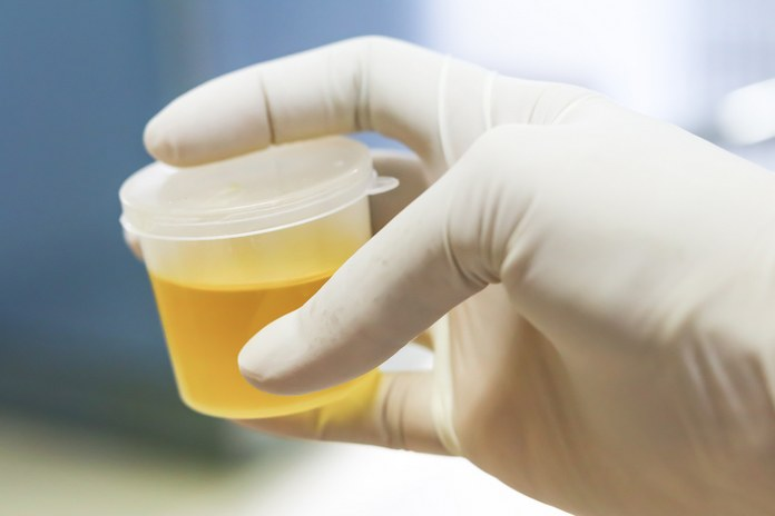
An examination of a urine specimen
Urine may be requested for identification of pathogens, red blood cells, or white blood cells. In order to avoid contaminating the urine sample, you may be asked to clean the genital area with an antiseptic pad before collecting the urine midstream.
Growing microorganisms from the urinary tract in a laboratory. Urine cultures are sometimes used following urine analysis in the laboratory. The results of this test are used to determine which microorganisms are responsible for the infection and which treatment is most likely to be effective.
Take photographs of your urinary tract. You can undergo an ultrasound, magnetic resonance imaging (MRI), or computerised tomography (CT) scan if your doctor suspects that an abnormality in your urinary tract is causing recurrent infections. Additionally, a contrast pigment is used to highlight structures within the urinary tract.
Utilisation of a scope to examine the bladder. If you have repeated UTIs, the doctor may conduct a cystoscopy. A long, thin tube with a lens (cystoscope) is used to view the urethra and bladder of a patient. The cystoscope is inserted into the bladder via the urethra.
Typically, urethritis is diagnosed through a thorough medical history (including social and sexual history) and physical examination. Patients with urethritis are typically healthy and exhibit no signs of systemic infection. The examination should concentrate on the pelvis, abdomen, and genitalia.
Men suffering from urethritis should undertake the following tests:
Penis: Examine for lesions that could indicate sexually transmitted diseases (such as herpes simplex, condyloma acuminatum, syphilis); in uncircumcised men, retract the foreskin to evaluate for lesions and fluid exudation.
Examine the lumen of the distal urethra for lesions, strictures, or obvious urethral discharges; palpate the urethra for tenderness, fluctuation, and warmth, which could indicate an abscess; and firmness, which could indicate a foreign body.
Examine the testicles for signs of inflammation or mass; feel the spermatic cord for enlargement, tenderness, or warmth, which may indicate orchitis or epididymitis.
Examine the patient for inguinal adenopathy.
Examine the prostate for tenderness or swelling that could indicate prostatitis.
Note any lesions in the perianal region during the digital rectal examination.
Observe female patients while they are in the lithotomy or frog-leg position. Include these assessments:
Examine lesions on the skin that may be indicative of other STDs.
The Urethra: Check for urethral discharge.
Pelvic region: pelvic examination, including the cervix
Common diagnostic procedures include:
Test for C-reactive protein
CBC stands for a complete blood count.
STI testing using a urine sample, whether for gonorrhoea or chlamydia.
Women are able to undergo pelvic ultrasounds.
The actions taken
Patients suffering from urethritis may require the following treatments:
In cases of urethral trauma, catheterization is performed to prevent tamponade urethral haemorrhage and urinary retention.
Cystoscopy: For catheter placement when catheterization is not possible; Extraction of extraneous objects or stones from the urethra
Amplatz dilators are utilised to dilate urethral strictures.
Suprapubic tube placement: In cases of severe urethral trauma where urethral catheterization is not possible or emergency cystoscopy facilities are unavailable; temporary measures to alleviate patient distress and divert urine.
