Diagnosis of Spider Vein

If you have spider veins, it is essential to see a doctor and have them properly diagnosed. In most instances, spider veins can be treated with simple procedures, but they can sometimes indicate a more severe condition. Your physician will be able to determine the cause of your spider veins and advise you on the most effective treatment. (7)
Examining the body
The best method to determine what causes spider veins and how to treat them is through a physical examination. The physician or surgeon will inspect the area for visible spider veins. They may also inquire about any additional symptoms you are experiencing and your family history of vascular disorders.
If the physician suspects that spider veins are the result of a more severe condition, he or she may order additional tests, such as an ultrasound or MRI.
Doppler sonography
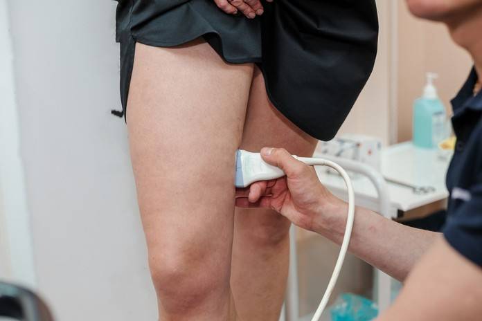
Ultrasound duplex venography is the most prevalent imaging technique. Doppler ultrasound is a noninvasive diagnostic instrument that creates images of blood vessels using sound waves. This technology can be used to identify spider veins and assess their severity. Doppler ultrasound enables physicians to observe blood flow through vessels, which can help them determine whether or not treatment is required.
The study of phlebography
Phlebography is an imaging technique used to detect veins and lymphatic vessels. It involves injecting a contrast agent into the vessels and obtaining x-ray or ultrasound images of them. This method is used to diagnose and treat vein diseases including varicose veins, spider veins, phlebitis, and lymphedema.
The Venogram
A venogram is a diagnostic procedure utilized to detect spider veins. It is a form of imaging test that creates an image of the body’s interior using x-rays. Contrast dye is injected into the veins before x-rays are obtained during a venogram. This allows the physician to determine the optimal course of treatment by determining whether the vessels are blocked.
