Evaluation of Osteoporosis

Osteoporosis is a disorder that steadily deteriorates bone health over time. If there is, then
A fissure that does not result from an external force.
In an elderly man, a woman after menopause, or a juvenile with malnutrition.
Soreness in the bones
General alterations to the physique and posture
Variations in locomotion and body structure
A family history of osteoporosis increases your risk of developing the disease.
Osteoporosis Diagnosis In The Laboratory

The most common and reliable test for confirming a diagnosis is the bone density test.
Bone density examination
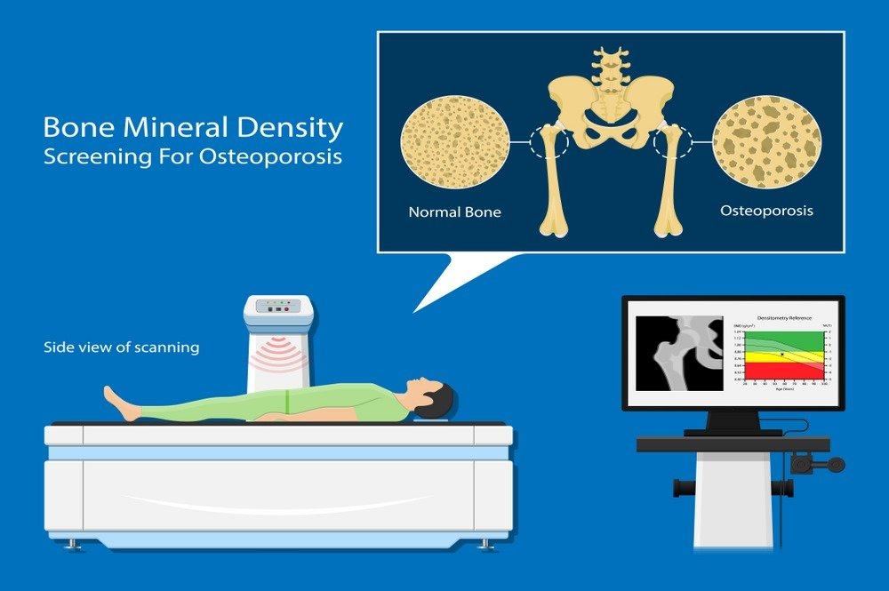
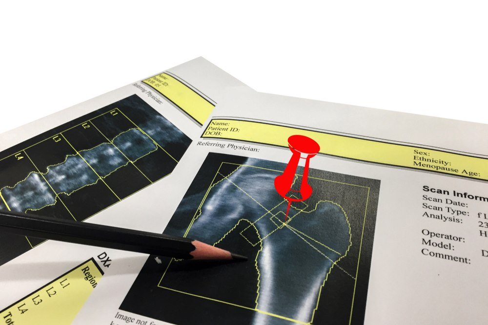
This noninvasive procedure can detect bone fragility prior to the onset of osteoporosis-related fractures. The axial skeleton is scanned using dual x-ray absorptiometry by a central DXA machine. It employs only low doses of radiation to generate an image of the bone and measure its mineralization. The hip or spine should be scanned first, as these areas are most susceptible to osteoporotic fractures. Normal x-rays can be used to determine the location of a fracture, but they are unable to detect bone density until the late phases of osteoporosis.
This test’s interpretation is based on the T-score, which compares the patient’s bone density to that of a normal 30-year-old.
A T-score greater than or equal to -1.0 indicates normal bone density.
A T-score between -1.0 and -2.5 indicates decreased bone density or the possibility of osteopenia.
A T-score of -2.5 or less indicates the presence of osteoporosis.
Based on the T-score value, the physician can determine next steps. It can help determine if the patient requires anti-osteoporosis medication or if simple lifestyle changes are sufficient to enhance bone health.
Test of Peripheral Bone Density

In the event that the central DXA is unavailable or cannot be performed on patients weighing more than 150 kilograms, the appendicular skeleton, including the wrist, digits, heel, and arm, can be evaluated. Use of the following procedures
(pDXA) peripheral dual x-ray absorptiometry
utilizing quantitative ultrasound (QUS)
pQCT is a peripheral quantitative computed tomography.
These exams are screening, not diagnostic, for osteoporosis. They only provide an estimate of bone density and mineralization and aid in determining the patient’s bone status. These tests provide a distinct indication of the need for additional tests.
FRAX Rating

A FRAX score document is a questionnaire that predicts the likelihood of fracture over the next decade. There is also a complimentary online service available. It considers the following factors that aid in evaluating bone density:
Age
Sex
Previous bone fracture history
Family ancestry
Size and Weight
Anti-inflammatory Glucocorticoid
cigarette smoking history
Alcohol consumption
Subsequent osteoporosis
The FRAX score is expressed as a percentage and is interpreted as a risk percentage or 10-year probability of two variables, the main osteoporotic fracture and the hip fracture.
Blood and Urine Examinations

The doctor may request blood and urine tests to identify any underlying condition that may cause bone loss. This includes calcium levels testing. The concentrations of reproductive hormones, thyroid, and parathyroid hormones are also measured in the samples.
