Evaluation of Ocular Migraine

When coping with ocular migraine, an accurate and timely diagnosis is essential. The first time a patient experiences a blind spot or transient vision loss, he should consult a physician. There is no definitive way to diagnose a migraine; however, the best approach is to rule out the possibility of any other eye disorder.
history of migraine headaches
During the consultation with an eye specialist, the physician will inquire about the patient’s family history of ocular migraine or classical migraine and general health. The likelihood of ocular migraine increases if the patient has a significant family history of the condition. Over fifty percent of individuals with ocular migraine have a family history. After being diagnosed with ocular migraine, 29% of patients complained of classical migraine. The physician will also ask about the frequency and severity of the symptoms. The probability that a person has ocular migraine increases with the frequency of their symptoms.
a fundoscopy
Fundus refers to the rear of the eye. It is made up of blood vessels, retina, optic disc, and macula. For observing the eye, the ophthalmoscope is a device with a lens and variable illumination settings. The affected eye’s blood vessels and retina will be observed using a variety of light intensities by the general practitioner. This procedure is non-painful and takes less than ten minutes. The ideal time to perform this examination is during an ocular migraine, so that the physician can examine the retina and vessels surrounding the eye in detail.
Throughout the method:
The patient is instructed to sit up straight while the doctor seats across from him or her.
The ophthalmoscope is held directly 6 inches from the eye, and the doctor adjusts the aperture in order to view various portions of the eye.
The patient is instructed to fixate on the object directly in front of them as the lights are dimmed.
In order to get a comprehensive view, the doctors may ask the patient to gaze in various directions.
Examination by slit-lamp
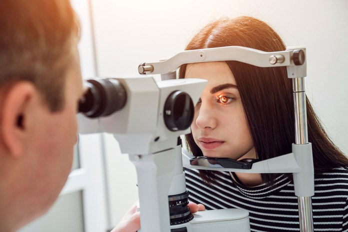
A slit lamp examination is another method for examining the eye, but it provides greater detail about the eye. The ophthalmologist can obtain a three-dimensional view of the eye during this examination. The doctor would investigate the patient’s eye using a microscope and light while the patient’s head is positioned in a frame. To dilate the pupil, the specialist may administer eye drops to the patient’s eye. The patient may also be asked to look in a variety of orientations in order to observe additional areas within the eye.
Various studies
If the doctor detects an abnormality, he may request that the patient undergo additional testing, such as a Computerised Testing scan or Magnetic Resonance Imaging. These tests are extremely useful for detecting haemorrhaging, tumours, and eye or head cancer. The physician may request that the patient undergo blood testing in order to identify any infection in the arteries and muscles effecting the eye or brain.
