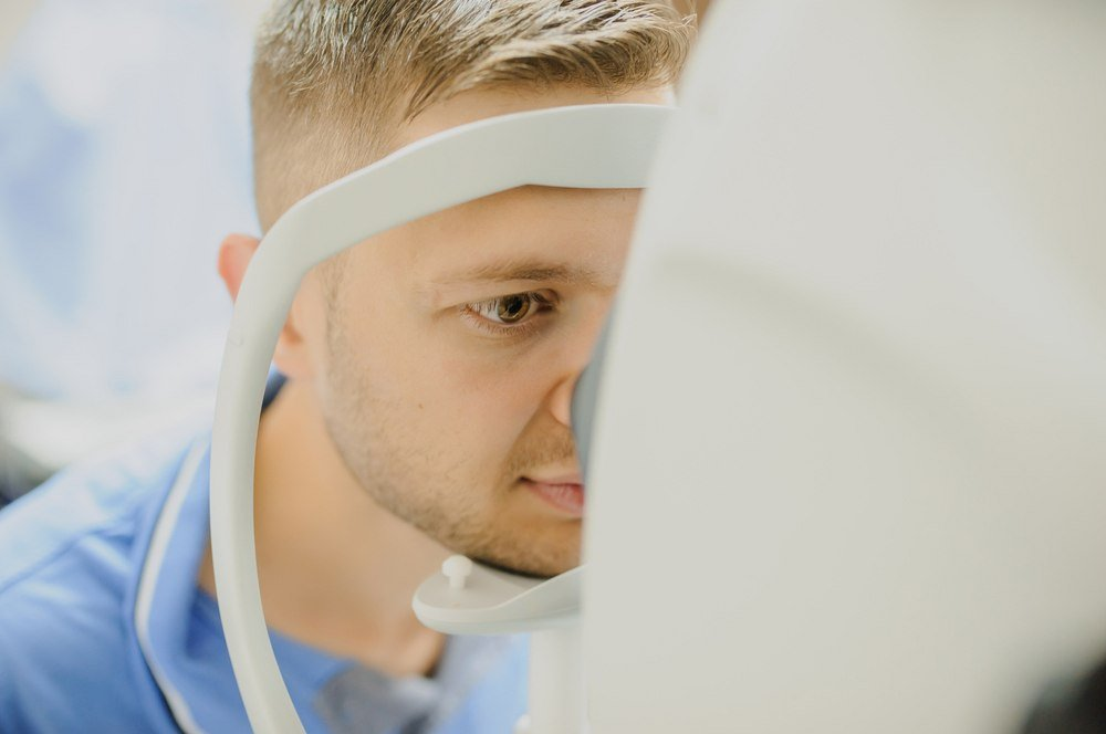
Imaging of Fundus Autofluorescence (FAF)

It is a noninvasive imaging technique that utilises the body’s natural fluorescence to examine the retina. When exposed to specific wavelengths of light, specific body structures illuminate. Retinal pigments, epithelial cells, and lipofuscin are examples of the fluorophores that make up this category. Typically, the procedure is used to study late dry AMD or geographic atrophy.
